

1.0
Cite Score 2025
SJR: 0.129 / SNIP: 0.140

JMERC
Owner
Janbazan Medical and Engineering Research Center (JMERC) is the owner of IJWPH.
Volume 17, Issue 4 (2025)
3 2025, 17(4): 383-389 |
Back to browse issues page
Request Type:




History
Received: 2025/12/13 | Accepted: 2025/12/22 | Published: 2025/11/19
Received: 2025/12/13 | Accepted: 2025/12/22 | Published: 2025/11/19
How to cite this article
Al-Rikabi H, Haji Ghasem Kashani M, Abiri E, Molaei Fard F, Altememy D. Effect of Tannic Acid on Apoptosis in the Testes of Mice Exposed to Mobile Phone Electromagnetic Waves. 3 2025; 17 (4) :383-389
URL: http://ijwph.daneshafarand.org/article-3-85661-en.html
URL: http://ijwph.daneshafarand.org/article-3-85661-en.html
Download citation:
BibTeX | RIS | EndNote | Medlars | ProCite | Reference Manager | RefWorks
Send citation to:


Rights and permissions
BibTeX | RIS | EndNote | Medlars | ProCite | Reference Manager | RefWorks
Send citation to:


Full-Text (HTML) (56 Views)
Introduction
The escalating use of radiofrequency wave generators, while providing numerous conveniences, also poses potential health risks to humans. Given the unavoidable nature of these devices, it is imperative to enhance our understanding of their mechanisms and develop strategies to minimize potential harm. The precise mechanisms by which mobile phone radiation impacts human health remain incompletely elucidated; however, two primary modes of action have been proposed [1].
Firstly, the “thermal effect” occurs at higher frequencies, wherein the generation of heat leads to an elevation in tissue or body temperature, subsequently resulting in the disruption of cellular function and development [2].
Secondly, the “non-thermal effect” involves the disruption of cellular membranes due to the passage of electrical impulses, leading to endothelial dysfunction, alterations in the blood-brain barrier, disturbances in cellular signal transduction, impacts on the immune system, and compromised excitability of the nervous system [1, 2].
The mechanism by which radiofrequency electromagnetic waves (RF-EMW) emitted by mobile phones operate likely involves a combination of both thermal and non-thermal effects [1, 2]. The reproductive system is among the organs susceptible to structural and functional disruptions from exposure to these waves, potentially culminating in infertility in both men and women [3]. The increasing personal use of mobile phones has raised concerns about the health implications of exposure to their radiation. Mobile phones emit low-frequency electromagnetic waves within the 800–2200MHz range, which are readily absorbed by the human body [4]. The energy absorbed by testicular tissue during electromagnetic exposure is readily converted to thermal energy, potentially elevating testicular temperature. This, in turn, can lead to a reduction in sperm count and motility, thereby contributing to infertility [3, 4].
Another study reports a significant decrease in protein kinase C and total sperm count, accompanied by an increase in apoptosis in male Wistar rats exposed to mobile phone radiation for 2 hours daily over 35 days [5]. It is generally established that mobile phone radiation can adversely affect fertility by disrupting the hypothalamic-pituitary-gonadal axis, inducing oxidative stress, and promoting apoptosis [6].
Apoptosis is executed through the activation of a family of cysteine proteases known as caspases. Caspases cleave proteins during the process of cellular demise [7, 8] and are categorized into three groups based on their function: initiator caspases, such as caspase 9; executioner caspases, such as caspase 3; and inflammatory caspases [9]. These caspases are initially expressed as inactive proenzymes. Upon activation, they participate in various stages of apoptosis. Apoptosis is the process by which cells die. It begins with the activation of initiator caspases, followed by executioner caspases [9]. Caspases are activated in response to a variety of stimuli, including DNA damage, growth factor deprivation, endoplasmic reticulum stress, and interactions with death receptors. This activation occurs through two primary pathways [10].
The extrinsic apoptosis pathway is initiated at the plasma membrane and involves death receptors, such as Fas, which are activated by extracellular ligands. Receptor activation leads to the formation of signaling complexes and the activation of caspase 8. Caspase 8 can activate executioner procaspases, such as caspase 3, and in some cases promote the release of cytochrome c via cleavage of the Bid protein, which subsequently activates caspase 9 and other executioner caspases [11, 12].
In the intrinsic apoptosis pathway, mitochondria play a central role and are activated in response to stress conditions. In this pathway, cytochrome c and other pro-apoptotic proteins are released from mitochondria into the cytosol. Cytochrome c combines with Apaf1 and dATP to form the apoptosome complex, which leads to the activation of caspase 3 and, subsequently, executioner caspases. Activated caspases induce nuclear fragmentation and the degradation of nuclear components. These two pathways not only possess distinct mechanisms but also converge on the ultimate activation of caspases and the execution of apoptosis [7, 13-15].
The apoptosome complex is formed by the oligomerization of Apaf1 in the presence of cytochrome c and ATP. Apoptosome formation is a critical step in the activation of caspase 9. In the presence of a strong apoptotic signal, the apoptosome complex is formed and poised to activate caspase 9, which provides a suitable environment for the activation of caspase 9 in the CARD domains [16, 17].
The Bcl-2 family members are key regulators of apoptosis via the intrinsic pathway [14]. Anti-apoptotic proteins, such as Bcl-2, are constitutively associated with intracellular membranes and prevent apoptosis. A high Bax/Bcl-2 ratio promotes apoptosis, and a decrease in Bcl-2 expression diminishes its inhibitory effect, leading to an increase in Bax expression and activation of caspase 3 [18]. BAD is a pro-apoptotic member of the Bcl-2 gene family that plays a role in the initiation of apoptosis. It has also been shown that BAD has many non-apoptotic functions that are closely related to cancer, including the regulation of glycolysis, autophagy, cell cycle progression, and the development of the body’s immune system. Unphosphorylated BAD forms a heterodimer with Bcl-2 and Bcl-xL, inactivating them and consequently leading to Bax/Bak-induced apoptosis. When BAD is phosphorylated by Akt/protein kinase B (which is activated by PIP3), it forms a 14-3-3-BAD protein heterodimer, causing Bcl-2 to be released to inhibit Bax-induced apoptosis; therefore BAD phosphorylation is anti-apoptotic, and BAD dephosphorylation is pro-apoptotic [19, 20].
The Fox family plays diverse roles during the development of organisms [21, 22]. FoxO proteins can induce apoptosis via both mitochondria-dependent intrinsic pathways and mitochondria-independent extrinsic pathways [23] and stimulate apoptosis by regulating the ratio of pro-apoptotic and survival factors (Bcl-2 family). They promote cell death by increasing the expression of pro-apoptotic factors and death receptor ligands, including TRAIL, FASL, Bim, and PUMA [24, 25].
Tannins are polyphenolic compounds classified into two main groups: hydrolyzable tannins (gallotannins and ellagitannins) and condensed tannins (containing flavonoid oligomers). Tannins exhibit significant medicinal properties, and plant extracts rich in tannins have demonstrated efficacy in clinical treatments, such as green tea, grape seeds, and polyphenol-rich chocolate [26].
Tannic acid (TA) is structurally the simplest hydrolyzable tannin, possessing the molecular formula C76H52O46, a glucose core, and 10 galloyl moieties [26]. TA exhibits antioxidant, antimicrobial, anti-inflammatory, and industrial applications and is effective in the treatment of diseases such as allergies, diabetes, and cardiovascular diseases. Furthermore, studies have shown that it possesses antitumor and anticancer properties by scavenging free radicals [27].
This study investigated the effect of TA on the expression of apoptosis-related genes, such as Bad, Apaf1, and Foxo3a, in the testicular tissue of mice exposed to mobile phone radiation.
Materials and Methods
In this experimental study, thirty 6-8-week-old adult male Balb/c mice were purchased from the Pasteur Institute of Tehran and housed for one week under controlled conditions of light (12 hours light/12 hours dark cycle) and temperature (20-22°C), with free access to water and standard laboratory chow.
The animals were randomly divided into five experimental groups. Control group consisted of mice that received no treatment. TA group consisted of mice that received TA at a dose of 20mg/kg intraperitoneally every other day (3 days per week). Sham group consisted of mice that received physiological saline instead of TA, with other conditions similar to the TA group. Electromagnetic field (EMF) group consisted of mice that were exposed to 900MHz electromagnetic waves for 8 hours daily (from 9:30 AM to 5:30 PM) [28]. TA+EMF group consisted of mice that were exposed to EMF as in the previous group and also received TA at a dose of 20mg/kg every other day (3 days per week). After 35 days of treatment, the animals were sacrificed, and the left testes were isolated and stored at -70°C for real-time polymerase chain reaction (PCR).
Total RNA was extracted from the left testes stored at -70°C using RNX-Plus, according to the manufacturer’s protocol. cDNA was then synthesized using the SinaClon company’s protocol.
Quantitative PCR (qPCR) was performed using RealQ Plus 2x Master Mix-Green Ampliqon, a fluorescent dye that binds to double-stranded DNA. The fluorescence signal was recorded after each reaction cycle by the instrument’s detector. The amount of fluorescence is proportional to the amount of PCR product generated in each cycle, indicating the amplification of the target DNA and gene of interest.
The target genes were normalized to the reference gene (EF1). The changes in relative gene expression were calculated using the Livak method:
ΔCt (Treated Group)=Ct (Target Gene)-Ct (Reference Gene)
ΔΔCt=ΔCt (Control)-ΔCt (Treated)
2-ΔΔCt = Livak Formula
qPCR reactions were performed using a Rotor-Gene 6000 series (Corbett) instrument and the reagents were as follows: cDNA: 0.5µL; SYBR Premix Ex Taq II: 5µL; forward primer: 0.5µL; reverse primer: 0.5µL; forward primer of the reference gene (EF1): 0.5µL; reverse primer of the reference gene (EF1): 0.5µL; DEPC water: 0.5µL; for a total volume of 20µL
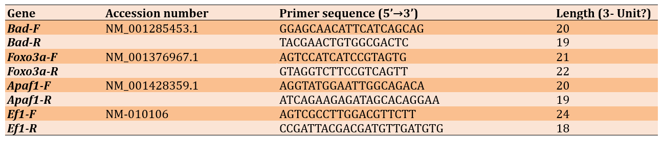
The target genes investigated were Foxo3a, Bad, and Apaf1. Primers for these genes were designed using Allele ID 7 software (Table 1).
Table 1. Sequence of primers used for quantitative PCR (qPCR)
The designed primers were synthesized by SinaClon Company and stored at -20°C after dilution. the reaction was performed according to the following program, with initial denaturation at 95°C for 10 minutes (1 cycle); denaturation at 95°C for 15 seconds (40 cycles); annealing at 60°C for 45 seconds (40 cycles); extension at 72°C for 30 seconds (40 cycles); and melting curve analysis at 60–95°C for 10 seconds (1 cycle).
Statistical analysis was performed using SPSS 16. To evaluate significant differences among the experimental groups, a one-way ANOVA was performed, followed by Tukey and LSD post hoc tests.
Findings
The average Bad gene expression level in the EMF group showed a significant increase compared to the other groups. A significant difference was also observed between the TA group and the sham group (p<0.1). No significant differences in Bad gene expression (p>0.05) were found among the other groups (Figure 1A). The average Apaf1 gene expression level in the EMF group showed a significant increase compared to the other groups. No significant differences in Apaf1 gene expression (p>0.05) were observed among the other groups (Figure 1B). The average Foxo3a gene expression level in the EMF group showed a significant increase compared to the other groups. Significant differences in Foxo3a gene expression were observed between the sham group and the EMF+TA and TA (20mg/kg) groups (p<0.001 and p<0.01, respectively). Significant differences in Foxo3a gene expression were also observed between the control group and the TA (20mg/kg) and EMF+TA groups (p<0.01 and p<0.1, respectively; Figure 1C).
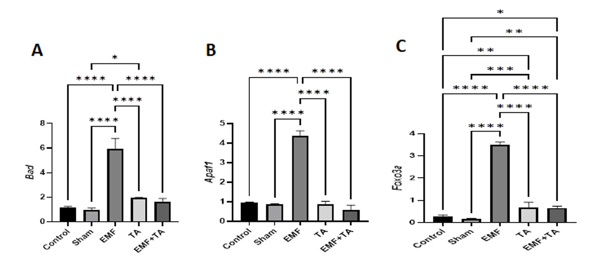
Figure 1. A) Changes in the expression levels of Bad in experimental groups; B) Changes in the expression levels of Apaf1 in experimental groups; C) Changes in the expression levels of Foxo3a in experimental groups.
Discussion
This study investigated the effect of TA on the expression of Bad, Apaf1, and Foxo3a in the testicular tissue of mice exposed to mobile phone radiation. Evidence suggests that exposure to radiofrequency radiation reduces testosterone levels, shrinks seminiferous tubules, increases apoptotic cells, and decreases sperm motility and count, thereby affecting testicular function. Exposure to 900MHz radiofrequency radiation emitted by mobile phones increased sperm caspase-3 activity, which can lead to apoptosis during spermatogenesis or sperm maturation and affect reproductive physiology. The data also suggest that decreased testosterone levels and abnormal sperm are potential causes of male infertility. These data are corroborated by increased reactive oxygen species (ROS) levels after exposure to radiofrequency radiation, as reported in previous studies, indicating an adverse effect on health [29].
In line with studies on the effects of magnetic waves on the testes, our study found that the group exposed to magnetic waves showed a significant increase in Apaf1 and Bad gene expression compared to the control group. Since these two genes are directly involved in the apoptosis pathway, this indicates that the apoptosis pathway was activated in the group exposed to magnetic waves. Furthermore, Foxo3a, which is indirectly involved in the apoptosis pathway, also showed significant expression in the EMF group, further indicating apoptosis activity in this group. The waves emitted from mobile phones are radiofrequency waves and are incapable of ionization or electrical stimulation, thus considered non-ionizing [19].
Radio waves originate from the movement of electrical charges in transmitting antennas and are emitted by the surface of these antennas. High-frequency radio waves are detrimental to the human body and lead to cancer and brain diseases [11].
Among the waves present in our environment, mobile phone waves pose the greatest risk to human health because mobile phones are in direct contact with the body. Moreover, adverse effects of excessive use of this device on humans manifest over the long term [30].
Numerous studies have investigated the effects of electromagnetic waves on tissue damage, yielding conflicting results. These discrepancies may arise from variations in tissue susceptibility, differing frequencies, magnetic flux densities, and varying exposure times across studies [12].
Damage to macromolecules, increased free radicals in cells, and ultimately DNA damage are among the detrimental effects of EMF exposure [11]. Studies examining the impact of mobile phone waves on the male reproductive system have reported alterations in testicular structure, disruption of spermatogenesis, decreased sperm motility, reduced Leydig cell numbers, and increased apoptosis of germ cells [31]. Kim et al. reported no significant adverse effects of mobile phones (150-1835MHz) on rat testes [30].
Houston et al. also demonstrated that short-term or intermittent exposure to 1800MHz radio-frequency radiation (RFR) has minor detrimental effects, whereas prolonged RFR exposure damages reproductive organs and sperm [32].
Al-Akhras et al. report that exposure of rats to a 50Hz, 25μT electromagnetic field significantly reduces total sperm count, slightly decreases FSH, increases LH, and significantly decreases testosterone levels [33].
Maclead was the first to report that ROS have detrimental effects on sperm and cause infertility. ROS can damage various biological molecules, including DNA, enzymes, lipids, and proteins [34]. A study by Kesari & Behari on the role of ROS in mobile phone exposure showed that ROS can also cause abnormal sperm production, increased caspase 3, and decreased testosterone in a group of aged Wistar rats [29].
A recent study reports increased DNA damage with elevated ROS, leading to histological and morphological changes in germ cells of Swiss albino mice exposed to 900MHz mobile phone radiation for 6 hours daily. Mitochondrial genome damage is also observed, and these DNA damages may lead to the accumulation of mutations and subsequently cancer development [28]. Prolonged exposure to radiation causes more severe DNA damage. However, short-term exposure has not been shown to cause genomic instability in studies. Nevertheless, it may affect the genome after several generations [35].
In a study by Kim et al., the effect of low-frequency electromagnetic fields on the apoptosis rate of male germ cells in mouse testes is investigated. The results showed that these waves have no significant effect on body and testicular histology, but the number of germ cell deaths is significantly increased in the experimental group compared to the control group [36]. A study found that electromagnetic fields increase free radical activity in cells. Studies have also shown that antioxidants prevent oxidative stress or apoptosis caused by electromagnetic fields in animal tissues [37].
Thus, EMF exerted its effect through the expression of these three apoptosis-related genes in the EMF group.
It has been reported that if the number of oxidants exceeds antioxidants, sperm may be affected primarily through three mechanisms: lipid peroxidation, DNA damage, and apoptosis induction. Antioxidants can potentially prevent the formation of excess ROS and repair cellular damage. Studies have also shown that TA can play a regulatory role in programmed cell death [38].
Experimental studies on TA have shown that intraperitoneal, intravenous, and even intranasal administration can achieve significant protective antioxidant, anti-inflammatory, and anti-apoptotic properties. TA can reduce ROS levels and decrease the expression of death receptor pathways and proteins related to the mitochondrial apoptosis signaling pathway, thereby preventing apoptosis in mice. Antioxidants are also essential for inhibiting EMF-related ROS production. Antioxidants prevent RF-EMF-induced apoptosis in various animal tissues [39].
We observed a significant decrease in Bad, Apaf1, and Foxo3a gene expression in the EMF+TA group compared to the EMF group, indicating the antioxidant effect of TA in inhibiting apoptosis in the EMF+TA group. However, we observed a significant difference in Bad gene expression between the TA and sham groups. Given that antioxidants can change their role at different doses and act as pro-oxidants [40], it is possible that this role reversal occurred in the TA group, leading to increased oxidative stress and consequently increased Bad gene expression compared to the sham group.
Furthermore, a significant difference in Foxo3a gene expression was observed between the TA group and the control and sham groups, which may again be due to the TA dose. Since Foxo3a has a dual role in stress response, its expression is moderate in low stress conditions, expressing Sod and Cat genes to combat stress. As stress increases, Foxo3a expression increases, expressing apoptosis-related genes [41].
Additionally, a significant difference in Foxo3a gene expression was observed between the EMF+TA group and the sham and control groups, which again relates to the dual role of Foxo3a. Since the control and sham groups experienced low stress, Foxo3a expression was lower in these groups compared to the EMF+TA group.
The EMF+TA group, being exposed to radiation and consequently experiencing high stress, should have shown high Foxo3a expression. However, administration of TA at 20mg/kg every other day prevented excessive expression of this apoptosis-related gene. The inability to reduce its expression to the levels of the sham and control groups is attributed to the high stress in this group. Perhaps by altering the dose or administering TA at shorter intervals, Foxo3a expression in this group could be brought closer to the levels of the sham and control groups.
The findings on Apaf1, Bad, and Foxo3a gene expression showed that EMF exposure affects the expression of apoptosis pathway genes, increasing their expression, which can induce apoptosis. TA, through its antioxidant properties, affected the radiation-exposed group and reduced the expression of apoptosis-related genes.
Conclusion
Tannic acid reduces apoptosis in the testes of mice exposed to radiofrequency electromagnetic fields, and exposure to 900MHz radiofrequency waves intensifies apoptosis in testicular tissue.
Acknowledgments: None declared.
Ethical Permissions: None declared.
Conflicts of Interests: The authors declared no conflicts of interests.
Authors' Contribution: Al-Rikabi HT (First Author), Introduction Writer/Methodologist/Main Researcher/Discussion Writer/Statistical Analyst (30%); Haji Ghasem Kashani M (Second Author), Introduction Writer/Methodologist/Assistant Researcher (20%); Abiri E (Third Author), Methodologist/Assistant Researcher/Discussion Writer (20%); Molaei Fard F (Fourth Author), Assistant Researcher/Discussion Writer/Statistical Analyst (20%); Altememy D (Fifth Author), Introduction Writer/Discussion Writer (10%)
Funding/Support: This project has received financial support from Damghan University, Damghan, Iran.
The escalating use of radiofrequency wave generators, while providing numerous conveniences, also poses potential health risks to humans. Given the unavoidable nature of these devices, it is imperative to enhance our understanding of their mechanisms and develop strategies to minimize potential harm. The precise mechanisms by which mobile phone radiation impacts human health remain incompletely elucidated; however, two primary modes of action have been proposed [1].
Firstly, the “thermal effect” occurs at higher frequencies, wherein the generation of heat leads to an elevation in tissue or body temperature, subsequently resulting in the disruption of cellular function and development [2].
Secondly, the “non-thermal effect” involves the disruption of cellular membranes due to the passage of electrical impulses, leading to endothelial dysfunction, alterations in the blood-brain barrier, disturbances in cellular signal transduction, impacts on the immune system, and compromised excitability of the nervous system [1, 2].
The mechanism by which radiofrequency electromagnetic waves (RF-EMW) emitted by mobile phones operate likely involves a combination of both thermal and non-thermal effects [1, 2]. The reproductive system is among the organs susceptible to structural and functional disruptions from exposure to these waves, potentially culminating in infertility in both men and women [3]. The increasing personal use of mobile phones has raised concerns about the health implications of exposure to their radiation. Mobile phones emit low-frequency electromagnetic waves within the 800–2200MHz range, which are readily absorbed by the human body [4]. The energy absorbed by testicular tissue during electromagnetic exposure is readily converted to thermal energy, potentially elevating testicular temperature. This, in turn, can lead to a reduction in sperm count and motility, thereby contributing to infertility [3, 4].
Another study reports a significant decrease in protein kinase C and total sperm count, accompanied by an increase in apoptosis in male Wistar rats exposed to mobile phone radiation for 2 hours daily over 35 days [5]. It is generally established that mobile phone radiation can adversely affect fertility by disrupting the hypothalamic-pituitary-gonadal axis, inducing oxidative stress, and promoting apoptosis [6].
Apoptosis is executed through the activation of a family of cysteine proteases known as caspases. Caspases cleave proteins during the process of cellular demise [7, 8] and are categorized into three groups based on their function: initiator caspases, such as caspase 9; executioner caspases, such as caspase 3; and inflammatory caspases [9]. These caspases are initially expressed as inactive proenzymes. Upon activation, they participate in various stages of apoptosis. Apoptosis is the process by which cells die. It begins with the activation of initiator caspases, followed by executioner caspases [9]. Caspases are activated in response to a variety of stimuli, including DNA damage, growth factor deprivation, endoplasmic reticulum stress, and interactions with death receptors. This activation occurs through two primary pathways [10].
The extrinsic apoptosis pathway is initiated at the plasma membrane and involves death receptors, such as Fas, which are activated by extracellular ligands. Receptor activation leads to the formation of signaling complexes and the activation of caspase 8. Caspase 8 can activate executioner procaspases, such as caspase 3, and in some cases promote the release of cytochrome c via cleavage of the Bid protein, which subsequently activates caspase 9 and other executioner caspases [11, 12].
In the intrinsic apoptosis pathway, mitochondria play a central role and are activated in response to stress conditions. In this pathway, cytochrome c and other pro-apoptotic proteins are released from mitochondria into the cytosol. Cytochrome c combines with Apaf1 and dATP to form the apoptosome complex, which leads to the activation of caspase 3 and, subsequently, executioner caspases. Activated caspases induce nuclear fragmentation and the degradation of nuclear components. These two pathways not only possess distinct mechanisms but also converge on the ultimate activation of caspases and the execution of apoptosis [7, 13-15].
The apoptosome complex is formed by the oligomerization of Apaf1 in the presence of cytochrome c and ATP. Apoptosome formation is a critical step in the activation of caspase 9. In the presence of a strong apoptotic signal, the apoptosome complex is formed and poised to activate caspase 9, which provides a suitable environment for the activation of caspase 9 in the CARD domains [16, 17].
The Bcl-2 family members are key regulators of apoptosis via the intrinsic pathway [14]. Anti-apoptotic proteins, such as Bcl-2, are constitutively associated with intracellular membranes and prevent apoptosis. A high Bax/Bcl-2 ratio promotes apoptosis, and a decrease in Bcl-2 expression diminishes its inhibitory effect, leading to an increase in Bax expression and activation of caspase 3 [18]. BAD is a pro-apoptotic member of the Bcl-2 gene family that plays a role in the initiation of apoptosis. It has also been shown that BAD has many non-apoptotic functions that are closely related to cancer, including the regulation of glycolysis, autophagy, cell cycle progression, and the development of the body’s immune system. Unphosphorylated BAD forms a heterodimer with Bcl-2 and Bcl-xL, inactivating them and consequently leading to Bax/Bak-induced apoptosis. When BAD is phosphorylated by Akt/protein kinase B (which is activated by PIP3), it forms a 14-3-3-BAD protein heterodimer, causing Bcl-2 to be released to inhibit Bax-induced apoptosis; therefore BAD phosphorylation is anti-apoptotic, and BAD dephosphorylation is pro-apoptotic [19, 20].
The Fox family plays diverse roles during the development of organisms [21, 22]. FoxO proteins can induce apoptosis via both mitochondria-dependent intrinsic pathways and mitochondria-independent extrinsic pathways [23] and stimulate apoptosis by regulating the ratio of pro-apoptotic and survival factors (Bcl-2 family). They promote cell death by increasing the expression of pro-apoptotic factors and death receptor ligands, including TRAIL, FASL, Bim, and PUMA [24, 25].
Tannins are polyphenolic compounds classified into two main groups: hydrolyzable tannins (gallotannins and ellagitannins) and condensed tannins (containing flavonoid oligomers). Tannins exhibit significant medicinal properties, and plant extracts rich in tannins have demonstrated efficacy in clinical treatments, such as green tea, grape seeds, and polyphenol-rich chocolate [26].
Tannic acid (TA) is structurally the simplest hydrolyzable tannin, possessing the molecular formula C76H52O46, a glucose core, and 10 galloyl moieties [26]. TA exhibits antioxidant, antimicrobial, anti-inflammatory, and industrial applications and is effective in the treatment of diseases such as allergies, diabetes, and cardiovascular diseases. Furthermore, studies have shown that it possesses antitumor and anticancer properties by scavenging free radicals [27].
This study investigated the effect of TA on the expression of apoptosis-related genes, such as Bad, Apaf1, and Foxo3a, in the testicular tissue of mice exposed to mobile phone radiation.
Materials and Methods
In this experimental study, thirty 6-8-week-old adult male Balb/c mice were purchased from the Pasteur Institute of Tehran and housed for one week under controlled conditions of light (12 hours light/12 hours dark cycle) and temperature (20-22°C), with free access to water and standard laboratory chow.
The animals were randomly divided into five experimental groups. Control group consisted of mice that received no treatment. TA group consisted of mice that received TA at a dose of 20mg/kg intraperitoneally every other day (3 days per week). Sham group consisted of mice that received physiological saline instead of TA, with other conditions similar to the TA group. Electromagnetic field (EMF) group consisted of mice that were exposed to 900MHz electromagnetic waves for 8 hours daily (from 9:30 AM to 5:30 PM) [28]. TA+EMF group consisted of mice that were exposed to EMF as in the previous group and also received TA at a dose of 20mg/kg every other day (3 days per week). After 35 days of treatment, the animals were sacrificed, and the left testes were isolated and stored at -70°C for real-time polymerase chain reaction (PCR).
Total RNA was extracted from the left testes stored at -70°C using RNX-Plus, according to the manufacturer’s protocol. cDNA was then synthesized using the SinaClon company’s protocol.
Quantitative PCR (qPCR) was performed using RealQ Plus 2x Master Mix-Green Ampliqon, a fluorescent dye that binds to double-stranded DNA. The fluorescence signal was recorded after each reaction cycle by the instrument’s detector. The amount of fluorescence is proportional to the amount of PCR product generated in each cycle, indicating the amplification of the target DNA and gene of interest.
The target genes were normalized to the reference gene (EF1). The changes in relative gene expression were calculated using the Livak method:
ΔCt (Treated Group)=Ct (Target Gene)-Ct (Reference Gene)
ΔΔCt=ΔCt (Control)-ΔCt (Treated)
2-ΔΔCt = Livak Formula
qPCR reactions were performed using a Rotor-Gene 6000 series (Corbett) instrument and the reagents were as follows: cDNA: 0.5µL; SYBR Premix Ex Taq II: 5µL; forward primer: 0.5µL; reverse primer: 0.5µL; forward primer of the reference gene (EF1): 0.5µL; reverse primer of the reference gene (EF1): 0.5µL; DEPC water: 0.5µL; for a total volume of 20µL
The target genes investigated were Foxo3a, Bad, and Apaf1. Primers for these genes were designed using Allele ID 7 software (Table 1).
Table 1. Sequence of primers used for quantitative PCR (qPCR)
The designed primers were synthesized by SinaClon Company and stored at -20°C after dilution. the reaction was performed according to the following program, with initial denaturation at 95°C for 10 minutes (1 cycle); denaturation at 95°C for 15 seconds (40 cycles); annealing at 60°C for 45 seconds (40 cycles); extension at 72°C for 30 seconds (40 cycles); and melting curve analysis at 60–95°C for 10 seconds (1 cycle).
Statistical analysis was performed using SPSS 16. To evaluate significant differences among the experimental groups, a one-way ANOVA was performed, followed by Tukey and LSD post hoc tests.
Findings
The average Bad gene expression level in the EMF group showed a significant increase compared to the other groups. A significant difference was also observed between the TA group and the sham group (p<0.1). No significant differences in Bad gene expression (p>0.05) were found among the other groups (Figure 1A). The average Apaf1 gene expression level in the EMF group showed a significant increase compared to the other groups. No significant differences in Apaf1 gene expression (p>0.05) were observed among the other groups (Figure 1B). The average Foxo3a gene expression level in the EMF group showed a significant increase compared to the other groups. Significant differences in Foxo3a gene expression were observed between the sham group and the EMF+TA and TA (20mg/kg) groups (p<0.001 and p<0.01, respectively). Significant differences in Foxo3a gene expression were also observed between the control group and the TA (20mg/kg) and EMF+TA groups (p<0.01 and p<0.1, respectively; Figure 1C).
Figure 1. A) Changes in the expression levels of Bad in experimental groups; B) Changes in the expression levels of Apaf1 in experimental groups; C) Changes in the expression levels of Foxo3a in experimental groups.
Discussion
This study investigated the effect of TA on the expression of Bad, Apaf1, and Foxo3a in the testicular tissue of mice exposed to mobile phone radiation. Evidence suggests that exposure to radiofrequency radiation reduces testosterone levels, shrinks seminiferous tubules, increases apoptotic cells, and decreases sperm motility and count, thereby affecting testicular function. Exposure to 900MHz radiofrequency radiation emitted by mobile phones increased sperm caspase-3 activity, which can lead to apoptosis during spermatogenesis or sperm maturation and affect reproductive physiology. The data also suggest that decreased testosterone levels and abnormal sperm are potential causes of male infertility. These data are corroborated by increased reactive oxygen species (ROS) levels after exposure to radiofrequency radiation, as reported in previous studies, indicating an adverse effect on health [29].
In line with studies on the effects of magnetic waves on the testes, our study found that the group exposed to magnetic waves showed a significant increase in Apaf1 and Bad gene expression compared to the control group. Since these two genes are directly involved in the apoptosis pathway, this indicates that the apoptosis pathway was activated in the group exposed to magnetic waves. Furthermore, Foxo3a, which is indirectly involved in the apoptosis pathway, also showed significant expression in the EMF group, further indicating apoptosis activity in this group. The waves emitted from mobile phones are radiofrequency waves and are incapable of ionization or electrical stimulation, thus considered non-ionizing [19].
Radio waves originate from the movement of electrical charges in transmitting antennas and are emitted by the surface of these antennas. High-frequency radio waves are detrimental to the human body and lead to cancer and brain diseases [11].
Among the waves present in our environment, mobile phone waves pose the greatest risk to human health because mobile phones are in direct contact with the body. Moreover, adverse effects of excessive use of this device on humans manifest over the long term [30].
Numerous studies have investigated the effects of electromagnetic waves on tissue damage, yielding conflicting results. These discrepancies may arise from variations in tissue susceptibility, differing frequencies, magnetic flux densities, and varying exposure times across studies [12].
Damage to macromolecules, increased free radicals in cells, and ultimately DNA damage are among the detrimental effects of EMF exposure [11]. Studies examining the impact of mobile phone waves on the male reproductive system have reported alterations in testicular structure, disruption of spermatogenesis, decreased sperm motility, reduced Leydig cell numbers, and increased apoptosis of germ cells [31]. Kim et al. reported no significant adverse effects of mobile phones (150-1835MHz) on rat testes [30].
Houston et al. also demonstrated that short-term or intermittent exposure to 1800MHz radio-frequency radiation (RFR) has minor detrimental effects, whereas prolonged RFR exposure damages reproductive organs and sperm [32].
Al-Akhras et al. report that exposure of rats to a 50Hz, 25μT electromagnetic field significantly reduces total sperm count, slightly decreases FSH, increases LH, and significantly decreases testosterone levels [33].
Maclead was the first to report that ROS have detrimental effects on sperm and cause infertility. ROS can damage various biological molecules, including DNA, enzymes, lipids, and proteins [34]. A study by Kesari & Behari on the role of ROS in mobile phone exposure showed that ROS can also cause abnormal sperm production, increased caspase 3, and decreased testosterone in a group of aged Wistar rats [29].
A recent study reports increased DNA damage with elevated ROS, leading to histological and morphological changes in germ cells of Swiss albino mice exposed to 900MHz mobile phone radiation for 6 hours daily. Mitochondrial genome damage is also observed, and these DNA damages may lead to the accumulation of mutations and subsequently cancer development [28]. Prolonged exposure to radiation causes more severe DNA damage. However, short-term exposure has not been shown to cause genomic instability in studies. Nevertheless, it may affect the genome after several generations [35].
In a study by Kim et al., the effect of low-frequency electromagnetic fields on the apoptosis rate of male germ cells in mouse testes is investigated. The results showed that these waves have no significant effect on body and testicular histology, but the number of germ cell deaths is significantly increased in the experimental group compared to the control group [36]. A study found that electromagnetic fields increase free radical activity in cells. Studies have also shown that antioxidants prevent oxidative stress or apoptosis caused by electromagnetic fields in animal tissues [37].
Thus, EMF exerted its effect through the expression of these three apoptosis-related genes in the EMF group.
It has been reported that if the number of oxidants exceeds antioxidants, sperm may be affected primarily through three mechanisms: lipid peroxidation, DNA damage, and apoptosis induction. Antioxidants can potentially prevent the formation of excess ROS and repair cellular damage. Studies have also shown that TA can play a regulatory role in programmed cell death [38].
Experimental studies on TA have shown that intraperitoneal, intravenous, and even intranasal administration can achieve significant protective antioxidant, anti-inflammatory, and anti-apoptotic properties. TA can reduce ROS levels and decrease the expression of death receptor pathways and proteins related to the mitochondrial apoptosis signaling pathway, thereby preventing apoptosis in mice. Antioxidants are also essential for inhibiting EMF-related ROS production. Antioxidants prevent RF-EMF-induced apoptosis in various animal tissues [39].
We observed a significant decrease in Bad, Apaf1, and Foxo3a gene expression in the EMF+TA group compared to the EMF group, indicating the antioxidant effect of TA in inhibiting apoptosis in the EMF+TA group. However, we observed a significant difference in Bad gene expression between the TA and sham groups. Given that antioxidants can change their role at different doses and act as pro-oxidants [40], it is possible that this role reversal occurred in the TA group, leading to increased oxidative stress and consequently increased Bad gene expression compared to the sham group.
Furthermore, a significant difference in Foxo3a gene expression was observed between the TA group and the control and sham groups, which may again be due to the TA dose. Since Foxo3a has a dual role in stress response, its expression is moderate in low stress conditions, expressing Sod and Cat genes to combat stress. As stress increases, Foxo3a expression increases, expressing apoptosis-related genes [41].
Additionally, a significant difference in Foxo3a gene expression was observed between the EMF+TA group and the sham and control groups, which again relates to the dual role of Foxo3a. Since the control and sham groups experienced low stress, Foxo3a expression was lower in these groups compared to the EMF+TA group.
The EMF+TA group, being exposed to radiation and consequently experiencing high stress, should have shown high Foxo3a expression. However, administration of TA at 20mg/kg every other day prevented excessive expression of this apoptosis-related gene. The inability to reduce its expression to the levels of the sham and control groups is attributed to the high stress in this group. Perhaps by altering the dose or administering TA at shorter intervals, Foxo3a expression in this group could be brought closer to the levels of the sham and control groups.
The findings on Apaf1, Bad, and Foxo3a gene expression showed that EMF exposure affects the expression of apoptosis pathway genes, increasing their expression, which can induce apoptosis. TA, through its antioxidant properties, affected the radiation-exposed group and reduced the expression of apoptosis-related genes.
Conclusion
Tannic acid reduces apoptosis in the testes of mice exposed to radiofrequency electromagnetic fields, and exposure to 900MHz radiofrequency waves intensifies apoptosis in testicular tissue.
Acknowledgments: None declared.
Ethical Permissions: None declared.
Conflicts of Interests: The authors declared no conflicts of interests.
Authors' Contribution: Al-Rikabi HT (First Author), Introduction Writer/Methodologist/Main Researcher/Discussion Writer/Statistical Analyst (30%); Haji Ghasem Kashani M (Second Author), Introduction Writer/Methodologist/Assistant Researcher (20%); Abiri E (Third Author), Methodologist/Assistant Researcher/Discussion Writer (20%); Molaei Fard F (Fourth Author), Assistant Researcher/Discussion Writer/Statistical Analyst (20%); Altememy D (Fifth Author), Introduction Writer/Discussion Writer (10%)
Funding/Support: This project has received financial support from Damghan University, Damghan, Iran.
Keywords:
References
1. WHO. WHO research agenda for radiofrequency fields. Geneva: World Health Organization; 2010. [Link]
2. Friedman J, Kraus S, Hauptman Y, Schiff Y, Seger R. Mechanism of short-term ERK activation by electromagnetic fields at mobile phone frequencies. Biochem J. 2007;405(3):559-68. [Link] [DOI:10.1042/BJ20061653]
3. La Vignera S, Condorelli RA, Vicari E, D'Agata R, Calogero AE. Effects of the exposure to mobile phones on male reproduction: A review of the literature. J Androl. 2012;33(3):350-6. [Link] [DOI:10.2164/jandrol.111.014373]
4. Agarwal A, Deepinder F, Sharma RK, Ranga G, Li J. Effect of cell phone usage on semen analysis in men attending infertility clinic: An observational study. Fertil Steril. 2008;89(1):124-8. [Link] [DOI:10.1016/j.fertnstert.2007.01.166]
5. Meo SA, Al-Drees AM, Husain S, Khan MM, Imran MB. Effects of mobile phone radiation on serum testosterone in Wistar albino rats. Saudi Med J. 2010;31(8):869-73. [Link] [DOI:10.15537/1658-3175.5093]
6. Sheiner EK, Sheiner E, Hammel RD, Potashnik G, Carel R. Effect of occupational exposures on male fertility: Literature review. Ind Health. 2003;41(2):55-62. [Link] [DOI:10.2486/indhealth.41.55]
7. Roshankhah S, Abdolmaleki A, Salahshoor MR. Anti-inflammatory, anti-apoptotic, and antioxidant actions of Middle Eastern Phoenix dactylifera extract on mercury-induced hepatotoxicity in vivo. Mol Biol Rep. 2020;47(8):6053-65. [Link] [DOI:10.1007/s11033-020-05680-4]
8. Gevrek F, Aydin D, Ozsoy S, Aygun H, Bicer C. Inhibition by Egb761 of the effect of cellphone radiation on the male reproductive system. Bratisl Lek Listy. 2017;118(11):676-83. [Link] [DOI:10.4149/BLL_2017_128]
9. Mancini M, Nicholson DW, Roy S, Thornberry NA, Peterson EP, Casciola-Rosen LA, et al. The caspase-3 precursor has a cytosolic and mitochondrial distribution: Implications for apoptotic signaling. J Cell Biol. 1998;140(6):1485-95. [Link] [DOI:10.1083/jcb.140.6.1485]
10. Kuwana T, Newmeyer DD. Bcl-2-family proteins and the role of mitochondria in apoptosis. Curr Opin Cell Biol. 2003;15(6):691-9. [Link] [DOI:10.1016/j.ceb.2003.10.004]
11. Mustafa M, Ahmad R, Tantry IQ, Ahmad W, Siddiqui S, Alam M, et al. Apoptosis: A comprehensive overview of signaling pathways, morphological changes, and physiological significance and therapeutic implications. Cells. 2024;13(22):1838. [Link] [DOI:10.3390/cells13221838]
12. Wajant H. The fas signaling pathway: More than a paradigm. Science. 2002;296(5573):1635-6. [Link] [DOI:10.1126/science.1071553]
13. Mobahat M, Narendran A, Riabowol K. Survivin as a preferential target for cancer therapy. Int J Mol Sci. 2014;15(2):2494-516. [Link] [DOI:10.3390/ijms15022494]
14. Ghasemian M, Mahdavi M, Zare P, Feizi MAH. Spiroquinazolinone-induced cytotoxicity and apoptosis in K562 human leukemia cells: Alteration in expression levels of Bcl-2 and Bax. J Toxicol Sci. 2015;40(1):115-26. [Link] [DOI:10.2131/jts.40.115]
15. Reed JC. Bcl-2-family proteins and hematologic malignancies: History and future prospects. Blood. 2008;111(7):3322-30. [Link] [DOI:10.1182/blood-2007-09-078162]
16. Riedl SJ, Salvesen GS. The apoptosome: Signalling platform of cell death. Nat Rev Mol Cell Biol. 2007;8(5):405-13. [Link] [DOI:10.1038/nrm2153]
17. Acehan D, Jiang X, Morgan DG, Heuser JE, Wang X, Akey CW. Three-dimensional structure of the apoptosome: Implications for assembly, procaspase-9 binding, and activation. Mol Cell. 2002;9(2):423-32. [Link] [DOI:10.1016/S1097-2765(02)00442-2]
18. Arbel N, Shoshan-Barmatz V. The anti-apoptotic protein Bcl2 regulates apoptosis via interaction with the mitochondrial protein, VDAC1. BIOCHIMICA ET BIOPHYSICA ACTA-BIOENERGETICS. 2010;1797:67. [Link] [DOI:10.1016/j.bbabio.2010.04.216]
19. Adachi M, Imai K. The proapoptotic BH3-only protein BAD transduces cell death signals independently of its interaction with Bcl-2. Cell Death Differ. 2002;9(11):1240-7. [Link] [DOI:10.1038/sj.cdd.4401097]
20. Pfeffer CM, Singh AT. Apoptosis: A target for anticancer therapy. Int J Mol Sci. 2018;19(2):448. [Link] [DOI:10.3390/ijms19020448]
21. Kume T, Deng K, Hogan BL. Murine forkhead/winged helix genes Foxc1 (Mf1) and Foxc2 (Mfh1) are required for the early organogenesis of the kidney and urinary tract. Development. 2000;127(7):1387-95. [Link] [DOI:10.1242/dev.127.7.1387]
22. Pelletier GJ, Brody SL, Liapis H, White RA, Hackett BP. A human forkhead/winged-helix transcription factor expressed in developing pulmonary and renal epithelium. Am J Physiol. 1998;274(3):L351-9. [Link] [DOI:10.1152/ajplung.1998.274.3.L351]
23. Arden KC. Multiple roles of FOXO transcription factors in mammalian cells point to multiple roles in cancer. Exp Gerontol. 2006;41(8):709-17. [Link] [DOI:10.1016/j.exger.2006.05.015]
24. Brunet A, Bonni A, Zigmond MJ, Lin MZ, Juo P, Hu LS, et al. Akt promotes cell survival by phosphorylating and inhibiting a Forkhead transcription factor. Cell. 1999;96(6):857-68. [Link] [DOI:10.1016/S0092-8674(00)80595-4]
25. Coomans De Brachène A, Demoulin JB. FOXO transcription factors in cancer development and therapy. Cell Mol Life Sci. 2016;73(6):1159-72. [Link] [DOI:10.1007/s00018-015-2112-y]
26. Jing W, Xiaolan C, Yu C, Feng Q, Haifeng Y. Pharmacological effects and mechanisms of tannic acid. Biomed Pharmacother. 2022;154:113561. [Link] [DOI:10.1016/j.biopha.2022.113561]
27. Kaczmarek B. Tannic acid with antiviral and antibacterial activity as a promising component of biomaterials-A minireview. Materials. 2020;13(14):3224. [Link] [DOI:10.3390/ma13143224]
28. Pandey N, Giri S, Das S, Upadhaya P. Radiofrequency radiation (900 MHz)-induced DNA damage and cell cycle arrest in testicular germ cells in Swiss albino mice. Toxicol Ind Health. 2017;33(4):373-84. [Link] [DOI:10.1177/0748233716671206]
29. Kesari KK, Behari J. Evidence for mobile phone radiation exposure effects on reproductive pattern of male rats: Role of ROS. Electromagn Biol Med. 2012;31(3):213-22. [Link] [DOI:10.3109/15368378.2012.700292]
30. Kim KH, Kabir E, Jahan SA. The use of cell phone and insight into its potential human health impacts. Environ Monit Assess. 2016;188(4):221. [Link] [DOI:10.1007/s10661-016-5227-1]
31. Roshangar L, Rad JS. Environmental electromagnetic field and female fertility. In: From preconception to postpartum. London: IntechOpen; 2012. [Link] [DOI:10.5772/30447]
32. Houston BJ, Nixon B, King BV, Aitken RJ, De Iuliis GN. Probing the origins of 1,800 MHz radio frequency electromagnetic radiation induced damage in mouse immortalized germ cells and spermatozoa in vitro. Front Public Health. 2018;6:270. [Link] [DOI:10.3389/fpubh.2018.00270]
33. Al-Akhras MA, Darmani H, Elbetieha A. Influence of 50 Hz magnetic field on sex hormones and other fertility parameters of adult male rats. Bioelectromagnetics. 2006;27(2):127-31. [Link] [DOI:10.1002/bem.20186]
34. MacLeod J. The role of oxygen in the metabolism and motility of human spermatozoa. Am J Physiol Leg Content. 1943;138(3):512-8. [Link] [DOI:10.1152/ajplegacy.1943.138.3.512]
35. Abdulkhaleq L, Assi M, Abdullah R, Zamri-Saad M, Taufiq-Yap Y, Hezmee M. The crucial roles of inflammatory mediators in inflammation: A review. Vet World. 2018;11(5):627-35. [Link] [DOI:10.14202/vetworld.2018.627-635]
36. Kim J, Ha CS, Lee HJ, Song K. Repetitive exposure to a 60-Hz time-varying magnetic field induces DNA double-strand breaks and apoptosis in human cells. Biochem Biophys Res Commun. 2010;400(4):739-44. [Link] [DOI:10.1016/j.bbrc.2010.08.140]
37. Desai NR, Kesari KK, Agarwal A. Pathophysiology of cell phone radiation: oxidative stress and carcinogenesis with focus on male reproductive system. Reprod Biol Endocrinol. 2009;7(1):114. [Link] [DOI:10.1186/1477-7827-7-114]
38. Pourmirzaei F, Ranjbaran M, Kadkhodaee M, Kianian F, Lorian K, Abdi A, et al. Sperm and testicular dysfunction during cecal ligation and puncture-induced sepsis in male rats and effects of tannic acid through reducing testicular oxidative stress and inflammation. Iran J Basic Med Sci. 2021;24(11):1554-60. [Link]
39. Nazıroğlu M, Yüksel M, Köse SA, Özkaya MO. Recent reports of Wi-Fi and mobile phone-induced radiation on oxidative stress and reproductive signaling pathways in females and males. J Membr Biol. 2013;246(12):869-75. [Link] [DOI:10.1007/s00232-013-9597-9]
40. Sotler R, Poljšak B, Dahmane R, Jukić T, Pavan Jukić D, Rotim C, et al. Prooxidant activities of antioxidants and their impact on health. Acta Clin Croat. 2019;58(4):726-36. [Link] [DOI:10.20471/acc.2019.58.04.20]
41. Pino E, Amamoto R, Zheng L, Cacquevel M, Sarria JC, Knott GW, et al. FOXO3 determines the accumulation of α-synuclein and controls the fate of dopaminergic neurons in the substantia nigra. Hum Mol Genet. 2014;23(6):1435-52. [Link] [DOI:10.1093/hmg/ddt530]