

1.0
Cite Score 2025
SJR: 0.129 / SNIP: 0.140

JMERC
Owner
Janbazan Medical and Engineering Research Center (JMERC) is the owner of IJWPH.
Volume 17, Issue 4 (2025)
3 2025, 17(4): 399-405 |
Back to browse issues page
Request Type:




History
Received: 2025/10/19 | Accepted: 2025/11/25 | Published: 2025/11/30
Received: 2025/10/19 | Accepted: 2025/11/25 | Published: 2025/11/30
How to cite this article
Rasheed Z, Ayyed H, Taha J. Assessment of Minimal Erythema Dose of Narrow Band Ultraviolet-B in a Sample of Iraqi Patients with Vitiligo and Psoriasis. 3 2025; 17 (4) :399-405
URL: http://ijwph.daneshafarand.org/article-3-85664-en.html
URL: http://ijwph.daneshafarand.org/article-3-85664-en.html
Download citation:
BibTeX | RIS | EndNote | Medlars | ProCite | Reference Manager | RefWorks
Send citation to:


Rights and permissions
BibTeX | RIS | EndNote | Medlars | ProCite | Reference Manager | RefWorks
Send citation to:


1- Department of Physics, Faculty of Education, Al-Iraqia University, Baghdad, Iraq
2- Department of Physiology, Faculty of Medicine, Al-Nahrain University, Baghdad, Iraq
2- Department of Physiology, Faculty of Medicine, Al-Nahrain University, Baghdad, Iraq
Full-Text (HTML) (54 Views)
Introduction
Ultraviolet (UV) light comprises a narrow band within the electromagnetic spectrum, with wavelengths between 100-400nm (UVC: 100-280nm; UVB: 280-320nm; UVA: 320-400nm) [1]. The Earth’s atmosphere absorbs UV light of shorter wavelengths, allowing only UV light of longer wavelengths (UVA, UVB, and a smaller amount of UVC) to reach the Earth’s surface [2]. UV light from the sun has been recognized as a treatment for both dermatological and systemic illnesses for centuries. Nevertheless, narrowband UVB (NBUVB) is a relatively current but effective addition to dermatological practice. In 1903, Dr. Niels Finsen conducted the first study on the use of UV radiation to treat skin diseases. Goeckerman followed in 1925 [3]. Phototherapy is a safe and effective therapeutic method that uses ultraviolet radiation (UVR) to treat specific diseases. Because UV has an immunosuppressive effect on cutaneous T cells and cytokines, it has been used in dermatology to treat a variety of skin conditions, including vitiligo, atopic dermatitis, and psoriasis [4]. UV phototherapy employs a specific spectrum of UV light, which can be divided into three irradiation ranges: Ultraviolet A (UVA), psoralen ultraviolet A (PUVA), and ultraviolet B (UVB). Broadband UVB (BB-UVB) at 280-320nm and NBUVB at 311-313nm are both included within the UVB category of phototherapy [5,6]. NBUVB has been demonstrated to have fewer adverse effects and to be more clinically tolerated than the previously discussed UV phototherapy [7, 8].
UV light from the sun or artificial sources has a significant immunosuppressive effect, partly mediated by inducing apoptosis in activated T cells. The immunosuppressive effects of UV light on the skin depend on several parameters, such as wavelength, radiation intensity, treatment dose, number of treatment sessions, and the optical properties of human skin. Generally, UVB light has a more significant immunosuppressive effect than UVA light. PUVA is a kind of photochemotherapy. Psoralens are small molecules that can enter cells and intercalate DNA. UVA light exposure can result in covalent binding of psoralens to DNA, leading to inhibition of cell proliferation and modification of the gene expression profile [9].
When choosing the appropriate treatment for patients, several factors, such as age, comorbidities, and disease severity, should be considered. Although there are various traditional biological drugs and agents for atopic dermatitis and psoriasis, phototherapeutic methods are still highly used for the treatment of inflammatory skin diseases.
Phototherapy is the use of UV radiation or visible light to treat various diseases. The roots of phototherapy date back to 1500 BC, when Hindus treated vitiligo, an autoimmune skin disease, using photosensitizing plant extracts and sun exposure. Natural sunlight (heliotherapy) has long been used for the treatment of various skin diseases, and it is still highly appealing for atopic dermatitis and psoriasis in several geographic areas in the world, especially in the Dead Sea [10]
UV-based phototherapy (including PUVA, UVA1, and UVB) has long been used successfully to treat many cutaneous disorders. Photoresponsive diseases have many causes; however, most involve disturbances in local and, occasionally, systemic inflammatory cells and/or abnormalities in keratinocytes, thereby triggering inflammation. UV-based phototherapy works through the regulation of the inflammatory component and causes apoptosis of pathogenic cells, resulting in a fascinating and complex network of simultaneous events, such as immediate transcriptional alterations in immune cells, keratinocytes, and pigment cells, the emergence of apoptotic bodies, and antigen-presenting cell trafficking in skin, which promptly transform the UV-exposed skin microenvironment. Recently, the mechanisms of UV-based phototherapy (PUVA, UVB, and UVA1) for many skin diseases, like pruritus, psoriasis, polymorphic light eruption (PLE), eczema, graft-versus-host disease (GvHD), fibrosing conditions, vitiligo, mast cell diseases, and cutaneous T-cell lymphoma (CTCL) have been unraveled. The integration of the immunomodulatory effects of phototherapy with the induction of apoptosis has been reported [11].
Two approaches are commonly used to plan NBUVB dosing. The first approach involves using preset dosages depending on the patient’s skin type; the most widely used classification is the Fitzpatrick skin type scale [12, 13]. The Fitzpatrick scale categorizes skin types. Type I always burns but never tans, type II always burns and sometimes tans, type III sometimes burns and always tans, type IV rarely burns and always tans, type V refers to moderately pigmented skin, often seen in Asian individuals, and type VI represents black skin, typical of individuals of African descent.
The second approach relies on determining each patient’s minimal erythema dose (MED), which reflects skin susceptibility to UV radiation from a specific light source and is defined as the lowest dose (J/cm²) required to produce minimal perceptible erythema at the site of skin phototesting [13, 14]. Visual detection of the MED is subjective and can be influenced by several parameters, including the light source, the dose increments used, the area exposed to radiation, and prior UV exposure [15]. After irradiation, erythema is typically assessed 24 hours later, two to three times per week. Therapy is initiated at 70% of the MED and increased in 20% increments. Subsequently, the dose is increased during successive sessions. This procedure is stopped when the patient’s skin becomes clear or nearly clear [16].
Determining the MED is crucial because NBUVB therapy, while effective for these conditions, carries risks of adverse effects like sunburn and phototoxicity if the dosage is not precisely controlled. Patient responses to phototherapy can vary significantly based on individual factors, including skin type, ethnicity, and the specific dermatological condition being treated. Iraqi patients, who often have skin types that fall within specific ranges of the Fitzpatrick scale (e.g., Types III-VI), may exhibit unique responses to NBUVB. Establishing a specific MED for this population ensures that treatment is both safe and effective, minimizing the risk of burns while maximizing therapeutic benefits. Without this targeted assessment, clinicians would rely on generalized NBUVB protocols, potentially leading to suboptimal treatment outcomes or increased adverse events for Iraqi patients with vitiligo and psoriasis. Therefore, this study is essential for tailoring NBUVB therapy to the specific needs of the local patient demographic, thereby optimizing treatment efficacy and patient safety.
This research aimed to determine the MED as a guide for dose calculation of NBUVB administered to patients with vitiligo and psoriasis treated at our center.
Materials and Methods
This analytical cross-sectional study was conducted among 21 patients with psoriasis and vitiligo who attended the Department of Dermatology at the Medical City Teaching Hospital in Baghdad over a four-month period. The sample included 13 females and 8 males, aged 9 to 60 years. Also, 15 patients had vitiligo, while 6 patients had psoriasis. Skin type was III in 10 patients and IV in 11 patients. The selected patients had no history of photosensitizing disorders, cancer, severe claustrophobia, segmental vitiligo, or spontaneous repigmentation.
Phototesting was carried out using a whole-body cabinet equipped with a UVB TL-01 Philips lamp emitting UV light at 311 nm. The device was set at a power of 2 milliwatts per square centimeter (mW/cm²) with an energy range of 200-600mJ/cm². Calibration of the UVB lamps was performed using a spectrophotometer traceable to national standards to ensure accurate irradiance measurement. Calibration was achieved by comparing the lamp output with a reference spectrophotometer of known spectral characteristics, allowing calculation of instrumental constants to convert measured voltages into irradiance values (W/m²) using the formula E=K×V, where E is the radiation value in W/m², V is the voltage measured by the meter in volts, and K is the calibration constant in W/m²/Volts.
The phototest was conducted on the forearm. The arm was covered with a black cloth with a 2×2 cm hole, while the rest of the body was also covered. NBUVB at a dose of 200mJ/cm² was used to irradiate the skin beneath the hole. The hole was then repositioned to expose another area, and the NBUVB dose was increased by 50mJ/cm² for each subsequent exposure. In total, six sites were irradiated with doses incrementally increased by 50mJ/cm² up to a maximum of 600mJ/cm². The selected dose range of 200-600mJ/cm² with 50mJ/cm² increments was based on scientific evidence from previous studies, manufacturers’ recommendations, and clinical considerations regarding patients’ skin sensitivity. This approach aimed to achieve a precise dose gradient, allowing for accurate and safe determination of the threshold for minimal skin redness (MED). The photo-tested sites were evaluated and photographed 24 hours later using a Sony 18.2‑megapixel digital camera (Figures 1 and 2).
A complete general, systemic, and dermatological examination was performed, taking into account the number of depigmented macules, the sites of involvement, and the approximate percentage of body surface area affected. This research was carried out in accordance with the ethical guidelines specified in the Declaration of Helsinki. Informed consent was obtained from all participants before their involvement in the study. The rights, confidentiality, and privacy of all participants were strictly protected throughout the research process. This research adhered to internationally accepted ethical standards for studies involving human subjects.
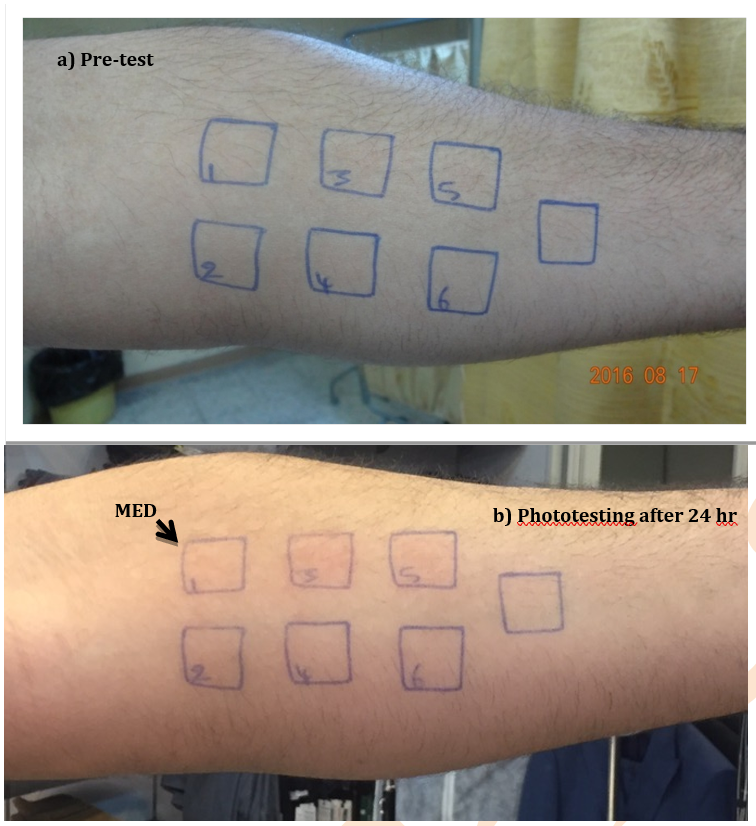
Figure 1. A 35-year-old male patient with vitiligo; a) Pre-test; b) 24 hours after the minimal erythema dose (MED) showing a response in the first patch (350mJ/cm²).
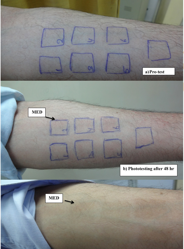
Figure 2. A 51-year-old male patient with psoriasis; a) Pre-test; b) 48 hours after the minimum erythema dose (MED) observed in the second patch (380mJ/cm2).
Data were analyzed using Microsoft Office Excel 2010 with a two-tailed t-test.
Findings
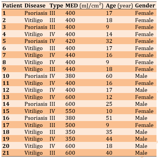
The mean MED and patients’ age were 448.1±87.6mJ/cm² (range: 350-600mJ/cm²) and 23.2±14.7 years, respectively (Table 1).
Table 1. Baseline characteristics and measured minimum erythema dose (MED) in patients with vitiligo and psoriasis
Some skin types appeared to have a wider range of MED values than others (Figure 3).
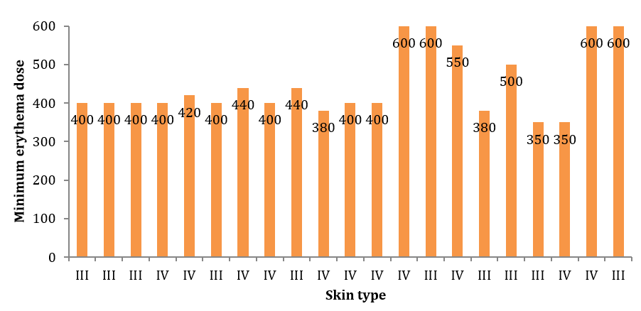
Figure 3. Relationship between minimum erythema dose (mJ/cm²) and skin type in the studied patients.
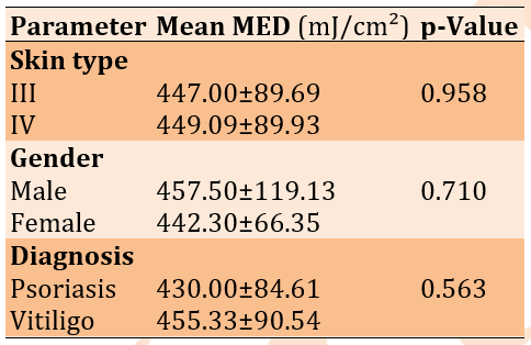
There were no statistically significant differences between males (457.50±119.13mJ/cm²) and females (442.30±66.35mJ/cm²; p=0.710). Likewise, no significant difference was observed between patients with psoriasis (430.00±84.61mJ/cm²) and those with vitiligo (455.33±90.5mJ/cm²; p=0.563). In addition, there was no significant difference between patients with skin type III (447.00±89.69mJ/cm²) and skin type IV (449.09±89.93mJ/cm²; p=0.958; Table 2).
Table 2. Comparison of mean minimal erythema dose (MED) by skin type, gender, and diagnosis
Discussion
This research aimed to determine MED among patients with vitiligo and psoriasis in Iraq. MED is a valuable tool for establishing dose regimens in the treatment of skin diseases using NBUVB. It is easy to perform and not time-consuming. The same device used to deliver NBUVB therapy can also be used to determine the MED. We demonstrated wide variation in MED values among patients, ranging from 350 to 600mJ/cm². However, no significant differences were observed between patients with psoriasis and those with vitiligo. Gender did not significantly affect MED, and no significant differences were found between patients with skin types III and IV.
Evaluation of skin sensitivity to UV radiation is an essential component of assessing cutaneous photosensitivity, particularly in conditions such as photodermatosis, photocarcinogenesis, sun protection, and phototherapy. One of the most commonly used measures of UV sensitivity is the MED, defined as the lowest dose of UV radiation that produces perceptible erythema on the skin. MED is typically assessed 24 hours after irradiation [17]. However, its visual assessment is subjective and may be influenced by several factors, including the light source, MED definition, irradiated area, dose increments, prior UV exposure, skin temperature, and inter- and intraobserver variability. Ethnic differences also contribute to variability, highlighting the need to validate MED measurements for specific populations.
UV sensitivity can also be assessed using indirect methods that do not require direct MED measurement. The most widely used approach is the Fitzpatrick skin type scale, although its reliability and validity have been questioned due to its subjective nature and susceptibility to recall bias. Despite these limitations, some authors suggest that the scale may be reliable if the evaluation process is standardized.
Studies conducted in various countries have examined the relationship between MED and skin type, with several reporting no consistent or favorable correlation between these parameters. In Colombia, the only published study, conducted among schoolchildren, similarly reports no correlation between UV-B MED and phototype [18]. The selection of the forearm for UV exposure and determination of the MED in Iraqi patients is based on several scientific and clinical considerations. It is well known that MED values vary between body regions due to differences in skin thickness, melanin content, and natural sun exposure. Areas, such as the face and hands, tend to be more sensitive than other regions, such as the back or forearm [14]. The forearm is a standard, commonly used site in clinical studies for MED determination because it is easily accessible, allows accurate dosing, facilitates monitoring of skin reactions, and is moderately exposed to sunlight, thereby reflecting an average skin sensitivity suitable for research purposes.
Skin sensitivity may vary in Iraqi patients due to ethnic and environmental factors. However, using the forearm provides a standardized site for comparing results locally and internationally, and allows for dose adjustments based on individual response [19]. Our results, which included Fitzpatrick skin types III and IV—the most common types in the Middle East—provide accurate indications for these categories. However, it also noted the presence of other skin types (I, II, V, and VI), which require consideration of their physical differences. Types I and II are considered to be more UV-sensitive and, therefore, may have a lower MED than types III and IV. This means that the doses used may have been high for these types, increasing the risk of burns or contributing to their occurrence.
Skin types V and VI are characterized by a higher melanin content than other types, providing greater natural protection and resulting in a higher MED; therefore, they may require higher doses to optimize therapeutic effect. Tejasvi et al. successfully determined the MED for narrowband ultraviolet B (NBUVB) radiation in North Indian patients by comparing visual assessments with derma spectrometer measurements. Their findings show no significant difference in MED as determined by visual and dermascope analyses. Patients with darker skin types do not show lower MED; however, some patients with lighter skin types do [20]. Jeon et al. conducted a study measuring the MED using spectrophotometric analysis following NBUVB phototesting and examined the clinical implications of these values for phototherapy. Among the Lab* color space parameters, only the b values showed a statistically significant difference between the skin type III and IV groups (p=0.003). A positive correlation was observed solely between MEDs and b values (p<0.05). Moreover, the mean b/L ratio was significantly higher in the type IV group compared with the type III group (p<0.05) [21].
Some studies report no association between MED and skin type [18], whereas others report an association [22]. Similar to narrowband UV-B, the average values for the Spanish population [23] (obtained by applying a conversion factor to UV-B MED values) are comparable to those reported by Valbuena et al. for all skin types [15]. In Detroit, USA, Carretero-Mangolis and Lim [24] declare that the median MED is 700mJ/cm2 for skin types III-IV and 600mJ/cm2 for skin types I-II. In 352 psoriasis patients in the UK, the median MED values by skin type were lower (200, 280, 390, and 550mJ/cm2 for skin types I–IV, respectively) [25].
The MED assessment is a valuable tool for customizing narrowband NBUVB phototherapy in patients with vitiligo and psoriasis. Future studies are necessary to strengthen the evidence supporting the use of MED assessment in customizing NBUVB phototherapy for patients with vitiligo and psoriasis. Large-scale, multicenter studies with more diverse patient populations are essential to validate the reproducibility and generalizability of MED-based dosing protocols across different ethnicities, skin phototypes, and disease clinical subtypes. Such research would help establish standardized guidelines that account for interindividual variability in UV sensitivity, thereby improving treatment precision and safety. In addition, longitudinal studies are needed to evaluate the long-term efficacy, safety profile, recurrence rates, and cumulative UV exposure risks associated with MED-guided NBUVB therapy. Comparative studies between MED-based dosing and conventional fixed-dose or phototype-based protocols would also clarify the clinical advantages and cost-effectiveness of individualized treatment strategies. Conducting this research is crucial to optimizing therapeutic outcomes, minimizing adverse effects, enhancing patient adherence, and ultimately improving the quality of life of individuals undergoing phototherapy.
Conclusion
While minimal erythema dose values vary across patients, disease status, gender, and skin types do not influence these measurements.
Acknowledgments: I would like to express my deepest gratitude and appreciation to all the medical and technical staff members of the Dermatology Department at Medical City Teaching Hospital, Baghdad, for their tremendous efforts and unwavering cooperation, which greatly contributed to the success of this research. Furthermore, I wish to extend my deepest thanks to the esteemed patients whose generous participation made it possible for us to carry out this study on a solid scientific foundation.
Ethical Permissions: The College of Education of Iraqi University has approved the ethical permission of this research (IRB-2024-06-DERM-001).
Conflicts of Interest: The authors declared no conflicts of interest.
Authors' Contribution: Rasheed ZA (First Author), Introduction Writer/Methodologist/Main Researcher/Discussion Writer (60%); Ayyed HK (Second Author), Introduction Writer/Statistical Analyst (20%); Taha JH (Third Author), Discussion Writer (20%)
Funding/Support: The research article didn’t receive any funding from any source (my university or any other institute).
Ultraviolet (UV) light comprises a narrow band within the electromagnetic spectrum, with wavelengths between 100-400nm (UVC: 100-280nm; UVB: 280-320nm; UVA: 320-400nm) [1]. The Earth’s atmosphere absorbs UV light of shorter wavelengths, allowing only UV light of longer wavelengths (UVA, UVB, and a smaller amount of UVC) to reach the Earth’s surface [2]. UV light from the sun has been recognized as a treatment for both dermatological and systemic illnesses for centuries. Nevertheless, narrowband UVB (NBUVB) is a relatively current but effective addition to dermatological practice. In 1903, Dr. Niels Finsen conducted the first study on the use of UV radiation to treat skin diseases. Goeckerman followed in 1925 [3]. Phototherapy is a safe and effective therapeutic method that uses ultraviolet radiation (UVR) to treat specific diseases. Because UV has an immunosuppressive effect on cutaneous T cells and cytokines, it has been used in dermatology to treat a variety of skin conditions, including vitiligo, atopic dermatitis, and psoriasis [4]. UV phototherapy employs a specific spectrum of UV light, which can be divided into three irradiation ranges: Ultraviolet A (UVA), psoralen ultraviolet A (PUVA), and ultraviolet B (UVB). Broadband UVB (BB-UVB) at 280-320nm and NBUVB at 311-313nm are both included within the UVB category of phototherapy [5,6]. NBUVB has been demonstrated to have fewer adverse effects and to be more clinically tolerated than the previously discussed UV phototherapy [7, 8].
UV light from the sun or artificial sources has a significant immunosuppressive effect, partly mediated by inducing apoptosis in activated T cells. The immunosuppressive effects of UV light on the skin depend on several parameters, such as wavelength, radiation intensity, treatment dose, number of treatment sessions, and the optical properties of human skin. Generally, UVB light has a more significant immunosuppressive effect than UVA light. PUVA is a kind of photochemotherapy. Psoralens are small molecules that can enter cells and intercalate DNA. UVA light exposure can result in covalent binding of psoralens to DNA, leading to inhibition of cell proliferation and modification of the gene expression profile [9].
When choosing the appropriate treatment for patients, several factors, such as age, comorbidities, and disease severity, should be considered. Although there are various traditional biological drugs and agents for atopic dermatitis and psoriasis, phototherapeutic methods are still highly used for the treatment of inflammatory skin diseases.
Phototherapy is the use of UV radiation or visible light to treat various diseases. The roots of phototherapy date back to 1500 BC, when Hindus treated vitiligo, an autoimmune skin disease, using photosensitizing plant extracts and sun exposure. Natural sunlight (heliotherapy) has long been used for the treatment of various skin diseases, and it is still highly appealing for atopic dermatitis and psoriasis in several geographic areas in the world, especially in the Dead Sea [10]
UV-based phototherapy (including PUVA, UVA1, and UVB) has long been used successfully to treat many cutaneous disorders. Photoresponsive diseases have many causes; however, most involve disturbances in local and, occasionally, systemic inflammatory cells and/or abnormalities in keratinocytes, thereby triggering inflammation. UV-based phototherapy works through the regulation of the inflammatory component and causes apoptosis of pathogenic cells, resulting in a fascinating and complex network of simultaneous events, such as immediate transcriptional alterations in immune cells, keratinocytes, and pigment cells, the emergence of apoptotic bodies, and antigen-presenting cell trafficking in skin, which promptly transform the UV-exposed skin microenvironment. Recently, the mechanisms of UV-based phototherapy (PUVA, UVB, and UVA1) for many skin diseases, like pruritus, psoriasis, polymorphic light eruption (PLE), eczema, graft-versus-host disease (GvHD), fibrosing conditions, vitiligo, mast cell diseases, and cutaneous T-cell lymphoma (CTCL) have been unraveled. The integration of the immunomodulatory effects of phototherapy with the induction of apoptosis has been reported [11].
Two approaches are commonly used to plan NBUVB dosing. The first approach involves using preset dosages depending on the patient’s skin type; the most widely used classification is the Fitzpatrick skin type scale [12, 13]. The Fitzpatrick scale categorizes skin types. Type I always burns but never tans, type II always burns and sometimes tans, type III sometimes burns and always tans, type IV rarely burns and always tans, type V refers to moderately pigmented skin, often seen in Asian individuals, and type VI represents black skin, typical of individuals of African descent.
The second approach relies on determining each patient’s minimal erythema dose (MED), which reflects skin susceptibility to UV radiation from a specific light source and is defined as the lowest dose (J/cm²) required to produce minimal perceptible erythema at the site of skin phototesting [13, 14]. Visual detection of the MED is subjective and can be influenced by several parameters, including the light source, the dose increments used, the area exposed to radiation, and prior UV exposure [15]. After irradiation, erythema is typically assessed 24 hours later, two to three times per week. Therapy is initiated at 70% of the MED and increased in 20% increments. Subsequently, the dose is increased during successive sessions. This procedure is stopped when the patient’s skin becomes clear or nearly clear [16].
Determining the MED is crucial because NBUVB therapy, while effective for these conditions, carries risks of adverse effects like sunburn and phototoxicity if the dosage is not precisely controlled. Patient responses to phototherapy can vary significantly based on individual factors, including skin type, ethnicity, and the specific dermatological condition being treated. Iraqi patients, who often have skin types that fall within specific ranges of the Fitzpatrick scale (e.g., Types III-VI), may exhibit unique responses to NBUVB. Establishing a specific MED for this population ensures that treatment is both safe and effective, minimizing the risk of burns while maximizing therapeutic benefits. Without this targeted assessment, clinicians would rely on generalized NBUVB protocols, potentially leading to suboptimal treatment outcomes or increased adverse events for Iraqi patients with vitiligo and psoriasis. Therefore, this study is essential for tailoring NBUVB therapy to the specific needs of the local patient demographic, thereby optimizing treatment efficacy and patient safety.
This research aimed to determine the MED as a guide for dose calculation of NBUVB administered to patients with vitiligo and psoriasis treated at our center.
Materials and Methods
This analytical cross-sectional study was conducted among 21 patients with psoriasis and vitiligo who attended the Department of Dermatology at the Medical City Teaching Hospital in Baghdad over a four-month period. The sample included 13 females and 8 males, aged 9 to 60 years. Also, 15 patients had vitiligo, while 6 patients had psoriasis. Skin type was III in 10 patients and IV in 11 patients. The selected patients had no history of photosensitizing disorders, cancer, severe claustrophobia, segmental vitiligo, or spontaneous repigmentation.
Phototesting was carried out using a whole-body cabinet equipped with a UVB TL-01 Philips lamp emitting UV light at 311 nm. The device was set at a power of 2 milliwatts per square centimeter (mW/cm²) with an energy range of 200-600mJ/cm². Calibration of the UVB lamps was performed using a spectrophotometer traceable to national standards to ensure accurate irradiance measurement. Calibration was achieved by comparing the lamp output with a reference spectrophotometer of known spectral characteristics, allowing calculation of instrumental constants to convert measured voltages into irradiance values (W/m²) using the formula E=K×V, where E is the radiation value in W/m², V is the voltage measured by the meter in volts, and K is the calibration constant in W/m²/Volts.
The phototest was conducted on the forearm. The arm was covered with a black cloth with a 2×2 cm hole, while the rest of the body was also covered. NBUVB at a dose of 200mJ/cm² was used to irradiate the skin beneath the hole. The hole was then repositioned to expose another area, and the NBUVB dose was increased by 50mJ/cm² for each subsequent exposure. In total, six sites were irradiated with doses incrementally increased by 50mJ/cm² up to a maximum of 600mJ/cm². The selected dose range of 200-600mJ/cm² with 50mJ/cm² increments was based on scientific evidence from previous studies, manufacturers’ recommendations, and clinical considerations regarding patients’ skin sensitivity. This approach aimed to achieve a precise dose gradient, allowing for accurate and safe determination of the threshold for minimal skin redness (MED). The photo-tested sites were evaluated and photographed 24 hours later using a Sony 18.2‑megapixel digital camera (Figures 1 and 2).
A complete general, systemic, and dermatological examination was performed, taking into account the number of depigmented macules, the sites of involvement, and the approximate percentage of body surface area affected. This research was carried out in accordance with the ethical guidelines specified in the Declaration of Helsinki. Informed consent was obtained from all participants before their involvement in the study. The rights, confidentiality, and privacy of all participants were strictly protected throughout the research process. This research adhered to internationally accepted ethical standards for studies involving human subjects.
Figure 1. A 35-year-old male patient with vitiligo; a) Pre-test; b) 24 hours after the minimal erythema dose (MED) showing a response in the first patch (350mJ/cm²).
Figure 2. A 51-year-old male patient with psoriasis; a) Pre-test; b) 48 hours after the minimum erythema dose (MED) observed in the second patch (380mJ/cm2).
Data were analyzed using Microsoft Office Excel 2010 with a two-tailed t-test.
Findings
The mean MED and patients’ age were 448.1±87.6mJ/cm² (range: 350-600mJ/cm²) and 23.2±14.7 years, respectively (Table 1).
Table 1. Baseline characteristics and measured minimum erythema dose (MED) in patients with vitiligo and psoriasis
Some skin types appeared to have a wider range of MED values than others (Figure 3).
Figure 3. Relationship between minimum erythema dose (mJ/cm²) and skin type in the studied patients.
There were no statistically significant differences between males (457.50±119.13mJ/cm²) and females (442.30±66.35mJ/cm²; p=0.710). Likewise, no significant difference was observed between patients with psoriasis (430.00±84.61mJ/cm²) and those with vitiligo (455.33±90.5mJ/cm²; p=0.563). In addition, there was no significant difference between patients with skin type III (447.00±89.69mJ/cm²) and skin type IV (449.09±89.93mJ/cm²; p=0.958; Table 2).
Table 2. Comparison of mean minimal erythema dose (MED) by skin type, gender, and diagnosis
Discussion
This research aimed to determine MED among patients with vitiligo and psoriasis in Iraq. MED is a valuable tool for establishing dose regimens in the treatment of skin diseases using NBUVB. It is easy to perform and not time-consuming. The same device used to deliver NBUVB therapy can also be used to determine the MED. We demonstrated wide variation in MED values among patients, ranging from 350 to 600mJ/cm². However, no significant differences were observed between patients with psoriasis and those with vitiligo. Gender did not significantly affect MED, and no significant differences were found between patients with skin types III and IV.
Evaluation of skin sensitivity to UV radiation is an essential component of assessing cutaneous photosensitivity, particularly in conditions such as photodermatosis, photocarcinogenesis, sun protection, and phototherapy. One of the most commonly used measures of UV sensitivity is the MED, defined as the lowest dose of UV radiation that produces perceptible erythema on the skin. MED is typically assessed 24 hours after irradiation [17]. However, its visual assessment is subjective and may be influenced by several factors, including the light source, MED definition, irradiated area, dose increments, prior UV exposure, skin temperature, and inter- and intraobserver variability. Ethnic differences also contribute to variability, highlighting the need to validate MED measurements for specific populations.
UV sensitivity can also be assessed using indirect methods that do not require direct MED measurement. The most widely used approach is the Fitzpatrick skin type scale, although its reliability and validity have been questioned due to its subjective nature and susceptibility to recall bias. Despite these limitations, some authors suggest that the scale may be reliable if the evaluation process is standardized.
Studies conducted in various countries have examined the relationship between MED and skin type, with several reporting no consistent or favorable correlation between these parameters. In Colombia, the only published study, conducted among schoolchildren, similarly reports no correlation between UV-B MED and phototype [18]. The selection of the forearm for UV exposure and determination of the MED in Iraqi patients is based on several scientific and clinical considerations. It is well known that MED values vary between body regions due to differences in skin thickness, melanin content, and natural sun exposure. Areas, such as the face and hands, tend to be more sensitive than other regions, such as the back or forearm [14]. The forearm is a standard, commonly used site in clinical studies for MED determination because it is easily accessible, allows accurate dosing, facilitates monitoring of skin reactions, and is moderately exposed to sunlight, thereby reflecting an average skin sensitivity suitable for research purposes.
Skin sensitivity may vary in Iraqi patients due to ethnic and environmental factors. However, using the forearm provides a standardized site for comparing results locally and internationally, and allows for dose adjustments based on individual response [19]. Our results, which included Fitzpatrick skin types III and IV—the most common types in the Middle East—provide accurate indications for these categories. However, it also noted the presence of other skin types (I, II, V, and VI), which require consideration of their physical differences. Types I and II are considered to be more UV-sensitive and, therefore, may have a lower MED than types III and IV. This means that the doses used may have been high for these types, increasing the risk of burns or contributing to their occurrence.
Skin types V and VI are characterized by a higher melanin content than other types, providing greater natural protection and resulting in a higher MED; therefore, they may require higher doses to optimize therapeutic effect. Tejasvi et al. successfully determined the MED for narrowband ultraviolet B (NBUVB) radiation in North Indian patients by comparing visual assessments with derma spectrometer measurements. Their findings show no significant difference in MED as determined by visual and dermascope analyses. Patients with darker skin types do not show lower MED; however, some patients with lighter skin types do [20]. Jeon et al. conducted a study measuring the MED using spectrophotometric analysis following NBUVB phototesting and examined the clinical implications of these values for phototherapy. Among the Lab* color space parameters, only the b values showed a statistically significant difference between the skin type III and IV groups (p=0.003). A positive correlation was observed solely between MEDs and b values (p<0.05). Moreover, the mean b/L ratio was significantly higher in the type IV group compared with the type III group (p<0.05) [21].
Some studies report no association between MED and skin type [18], whereas others report an association [22]. Similar to narrowband UV-B, the average values for the Spanish population [23] (obtained by applying a conversion factor to UV-B MED values) are comparable to those reported by Valbuena et al. for all skin types [15]. In Detroit, USA, Carretero-Mangolis and Lim [24] declare that the median MED is 700mJ/cm2 for skin types III-IV and 600mJ/cm2 for skin types I-II. In 352 psoriasis patients in the UK, the median MED values by skin type were lower (200, 280, 390, and 550mJ/cm2 for skin types I–IV, respectively) [25].
The MED assessment is a valuable tool for customizing narrowband NBUVB phototherapy in patients with vitiligo and psoriasis. Future studies are necessary to strengthen the evidence supporting the use of MED assessment in customizing NBUVB phototherapy for patients with vitiligo and psoriasis. Large-scale, multicenter studies with more diverse patient populations are essential to validate the reproducibility and generalizability of MED-based dosing protocols across different ethnicities, skin phototypes, and disease clinical subtypes. Such research would help establish standardized guidelines that account for interindividual variability in UV sensitivity, thereby improving treatment precision and safety. In addition, longitudinal studies are needed to evaluate the long-term efficacy, safety profile, recurrence rates, and cumulative UV exposure risks associated with MED-guided NBUVB therapy. Comparative studies between MED-based dosing and conventional fixed-dose or phototype-based protocols would also clarify the clinical advantages and cost-effectiveness of individualized treatment strategies. Conducting this research is crucial to optimizing therapeutic outcomes, minimizing adverse effects, enhancing patient adherence, and ultimately improving the quality of life of individuals undergoing phototherapy.
Conclusion
While minimal erythema dose values vary across patients, disease status, gender, and skin types do not influence these measurements.
Acknowledgments: I would like to express my deepest gratitude and appreciation to all the medical and technical staff members of the Dermatology Department at Medical City Teaching Hospital, Baghdad, for their tremendous efforts and unwavering cooperation, which greatly contributed to the success of this research. Furthermore, I wish to extend my deepest thanks to the esteemed patients whose generous participation made it possible for us to carry out this study on a solid scientific foundation.
Ethical Permissions: The College of Education of Iraqi University has approved the ethical permission of this research (IRB-2024-06-DERM-001).
Conflicts of Interest: The authors declared no conflicts of interest.
Authors' Contribution: Rasheed ZA (First Author), Introduction Writer/Methodologist/Main Researcher/Discussion Writer (60%); Ayyed HK (Second Author), Introduction Writer/Statistical Analyst (20%); Taha JH (Third Author), Discussion Writer (20%)
Funding/Support: The research article didn’t receive any funding from any source (my university or any other institute).
References
1. Hamouda SA, Alshawish NK, Abdalla YK, Ibrahim MK. Ultraviolet radiation: Health risks and benefits. Saudi J Eng Technol. 2022;7(10):533-41. [Link] [DOI:10.36348/sjet.2022.v07i10.001]
2. Chilingar GV, Sorokhtin OG, Khilyuk LF, Haroun M, Albannay A. Absorption of the solar ultraviolet radiation by the earth's atmosphere and ozone formation. J Sustain Energy Eng. 2013;1(2):161-8. [Link] [DOI:10.7569/JSEE.2012.629509]
3. Myers E, Kheradmand S, Miller R. An update on narrowband ultraviolet B therapy for the treatment of skin diseases. Cureus. 2021;13(11):e19182. [Link] [DOI:10.7759/cureus.19182]
4. Salman KE, Altunay I, Salman A. The efficacy and safety of targeted narrowband UVB therapy: A retrospective cohort study. Turk J Med Sci. 2019;49(2):595-603. [Link] [DOI:10.3906/sag-1810-110]
5. Brenner AV, Lubin JH, Calista D, Landi MT. Instrumental measurements of skin color and skin ultraviolet light sensitivity and risk of cutaneous malignant melanoma: A case-control study in an Italian population. Am J Epidemiol. 2002;156(4):353-62. [Link] [DOI:10.1093/aje/kwf045]
6. Kaur HS, Chen JS, Doolan BJ, Gupta M. Narrow-band UVB phototherapy-Australian consensus-based recommendations. Australas J Dermatol. 2022;63(3):387-91. [Link] [DOI:10.1111/ajd.13850]
7. Huang MW, Lo PY, Cheng KS. Objective assessment of sunburn and minimal erythema doses: Comparison of noninvasive in vivo measuring techniques after UVB irradiation. EURASIP J Adv Signal Process. 2010;2010:1-7. [Link] [DOI:10.1155/2010/483562]
8. Ibbotson SH. A perspective on the use of NB-UVB phototherapy vs. PUVA photochemotherapy. Front Med. 2018;5:184. [Link] [DOI:10.3389/fmed.2018.00184]
9. Derheimer FA, Hicks JK, Paulsen MT, et al. Psoralen-induced DNA interstrand cross-links block transcription and induce p53 in an ataxia-telangiectasia and rad3-related-dependent manner. Mol Pharmacol. 2009;75:599-607. [Link] [DOI:10.1124/mol.108.051698]
10. Matos TR, Ling TC, Sheth V. Ultraviolet B radiation therapy for psoriasis: pursuing the optimal regime. Clin Dermatol. 2016;34:587-593. [Link] [DOI:10.1016/j.clindermatol.2016.05.008]
11. Vieyra-Garcia PA, Wolf P. A deep dive into UV-based phototherapy: Mechanisms of action and emerging molecular targets in inflammation and cancer. Pharmacol Ther. 2021;222: 107784. [Link] [DOI:10.1016/j.pharmthera.2020.107784]
12. Mai SN, Pham GP, Van Toi V, Huynh K, Pham TTH. Using UVB 311 nm narrow-band medical lamp for the treatment of psoriasis. Proceedings of the 7th International Conference on the Development of Biomedical Engineering in Vietnam (BME7) Translational Health Science and Technology for Developing Countries 7. Singapore: Springer; 2020. p. 413-9. [Link] [DOI:10.1007/978-981-13-5859-3_73]
13. Almokhtar MN. Phototesting for a variety of skin conditions before narrowband (TL-01) Ultraviolet-B. Grn Int J Apl Med Sci. 2024;2(5):182-8. [Link] [DOI:10.62046/gijams.2024.v02i05.004]
14. Lee EJ, Lee CH, Baek JH, Koh JS, Boo YC. A spectrophotometric method to determine minimal erythema dose for ultraviolet radiation in human skin. Biomedicines. 2024;12(11):1-15. [Link] [DOI:10.3390/biomedicines12112544]
15. Valbuena Mesa MC, Nova Villanueva JA, Sánchez Vanegas G. Minimal erythema dose: Correlation with Fitzpatrick skin type and concordance between methods of erythema assessment in a patient sample in Colombia. Actas Dermosifiliogr. 2020;111(5):390-7. [Link] [DOI:10.1016/j.adengl.2019.12.010]
16. Dogra S, De D. Narrowband ultraviolet B in the treatment of psoriasis: The journey so far!. Indian J Dermatol Venereol Leprol. 2010;76(6):652-61. [Link] [DOI:10.4103/0378-6323.72461]
17. Graham IH, Antony RY. Ultraviolet radiation-induced erythema in human skin. Methods. 2002; 28(1): 14. [Link] [DOI:10.1016/S1046-2023(02)00205-0]
18. Sanclemente G, Zapata JF, J.J. García, A. Gaviria, L.F. Gómez, M. Barrera. Lack of correlation between minimal erythema dose and skin phototype in a Colombian scholar population. Skin Res Technol. 2008;14:403-409. [Link] [DOI:10.1111/j.1600-0846.2008.00306.x]
19. Yang JW, Fan GB, Tan F, Kong HM, Liu Q, Zou Y, et al. The role and safety of UVA and UVB in UV-induced skin erythema. Front Med. 2023;10:1163697. [Link] [DOI:10.3389/fmed.2023.1163697]
20. Tejasvi T, Sharma VK, Kaur J. Determination of minimal erythemal dose for narrow band-ultraviolet B radiation in north Indian patients: Comparison of visual and dermaspectrometer readings. Indian J Dermatol Venereol Leprol. 2007;73(2):97-9. [Link] [DOI:10.4103/0378-6323.31893]
21. Jeon SY, Lee CY, Song KH, Kim KH. Spectrophotometric measurement of minimal erythema dose sites after narrowband ultraviolet B phototesting: Clinical implication of spetrophotometric values in phototherapy. Ann Dermatol. 2014;26(1):17-25. [Link] [DOI:10.5021/ad.2014.26.1.17]
22. Li YW, Chu CY. The minimal erythema dose of broadband ultraviolet B in Taiwanese. J Formos Med Assoc. 2007;106:975-978. [Link] [DOI:10.1016/S0929-6646(08)60071-6]
23. Pérez Ferriols A, Aguilera J, Aguilera P, de Argila D, Barnadas MA, de Cabo X, et al. Determination of minimal erythema dose and anomalous reactions to UVA radiation by skin phototype. Actas Dermosifiliogr. 2014; 105:780-788. [Link] [DOI:10.1016/j.adengl.2014.05.020]
24. Carretero-Mangolis C, Lim HW. Correlation between skin types and minimal erythema dose in narrowband UVB (TL-01) phototherapy. Photodermatol Photoimmunol Photomed. 2001;17:244-246. [Link] [DOI:10.1111/j.1600-0781.2001.170508.x]
25. Palmer RA, Aquilina S, Milligan PJ, Walker SL, Hawk JL, Young AR. Photoadaptation during narrowband ultraviolet-B therapy is independent of skin type: a study of 352 patients. J Invest Dermatol. 2006;126:1256-1263, [Link] [DOI:10.1038/sj.jid.5700287]